It's not just what we know... it's how much we care!
Ultrasonography & Radiography
At Wingham and Valley Vets we know that accurate diagnosis is the cornerstone of good quality medicine. Our ultrasonography service and top-quality ultrasound equipment allow us to routinely obtain diagnoses which previously required referral to a specialist centre, or invasive exploratory surgery.
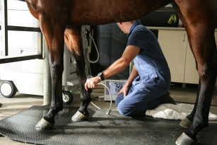
Ultrasound is an indispensable tool to diagnose many conditions of large and small animals, particularly those involving soft tissues, such as organs in the abdomen or chest. A full abdominal ultrasound yields a huge amount of information about your pet’s health. Even a ‘normal’ abdominal ultrasound scan is highly useful, as it rules out a multitude of potentially life-threatening conditions.
Several of our vets have a particular interest in ultrasonography and Michael Healy has undertaken further study in ultrasound, performing ultrasounds of the abdomen, thoracic cavity and musculoskeletal system.
What is an ultrasound scan?

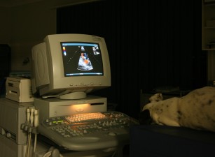
An echocardiogram – ultrasound scan of the heart.
Ultrasound scanning uses high frequency sound waves to produce images of structures within the body. Some sound waves are absorbed by body tissues and others bounce back. The ultrasound machine measures the sound waves that bounce back and transforms them into an image on a screen. Extensive training is required in order to correctly use this equipment and interpret these images. Ultrasound is most useful for looking at soft-tissue structures and organs like the liver, kidney, intestines, bladder and heart. It is less effective for examining bones (unlike x-rays).
What happens when MY PET is booked in for an ultrasound?
If it is an emergency ultrasound, we will do it immediately.
For routine ultrasounds we will book an appointment and depending on the nature of their symptoms, may admit them to hospital for the day. Since ultrasound is a painless procedure, most animals will allow a full examination without sedation, particularly if the owner can stay for the procedure. However, you should bring your pet in fasted on the morning of admission, as they may need to be sedated to ensure we can get high quality images. In addition, special intestinal tract scans may require 24 hours of fasting.
Usually your pet will need to be shaved over the area that is being scanned. During the scan a gel is applied over the clipped area to be examined and a transducer (probe) is placed on the skin.
Once the scan has been done we will give you a call or book an appointment for our veterinarians to show you the images and to discuss the diagnosis and treatment plan for your pet. However if you wish, you may be present during your pet’s ultrasound.
WHY MIGHT MY PET NEED AN ULTRASOUND?
- Pregnancy ultrasounds to confirm pregnancy and assess foetal viability as well as for the management of ‘high risk’ pregnancies (in conjunction with progesterone monitoring at breeders’ request).
- Gastrointestinal problems such as chronic vomiting, diarrhoea, inappetance, abdominal pain and unexplained weight loss.
- Heart problems such as murmurs or arrhythmias.
- Urinary issues such as blood in the urine, painful urination or increased frequency.
- Emergencies such as car accidents, dog attacks, blunt force trauma, sudden collapse etc.

If you have any further questions about our ultrasonography service, please contact the clinic on 6557 0000.
At Wingham and Valley Vets we know that accurate diagnosis is the cornerstone of good quality medicine. Our ultrasonography service and top-quality ultrasound equipment allow us to routinely obtain diagnoses which previously required referral to a specialist centre, or invasive exploratory surgery.
Ultrasound is an indispensable tool to diagnose many conditions of large and small animals, particularly those involving soft tissues, such as organs in the abdomen or chest. A full abdominal ultrasound yields a huge amount of information about your pet’s health. Even a ‘normal’ abdominal ultrasound scan is highly useful, as it rules out a multitude of potentially life-threatening conditions.
Several of our vets have a particular interest in ultrasonography and Michael Healy has undertaken further study in ultrasound, performing ultrasounds of the abdomen, thoracic cavity and musculoskeletal system.
What is an ultrasound scan?

An echocardiogram – ultrasound scan of the heart.
Ultrasound scanning uses high frequency sound waves to produce images of structures within the body. Some sound waves are absorbed by body tissues and others bounce back. The ultrasound machine measures the sound waves that bounce back and transforms them into an image on a screen. Extensive training is required in order to correctly use this equipment and interpret these images. Ultrasound is most useful for looking at soft-tissue structures and organs like the liver, kidney, intestines, bladder and heart. It is less effective for examining bones (unlike x-rays).
What happens when MY PET is booked in for an ultrasound?
If it is an emergency ultrasound, we will do it immediately.
For routine ultrasounds we will book an appointment and depending on the nature of their symptoms, may admit them to hospital for the day. Since ultrasound is a painless procedure, most animals will allow a full examination without sedation, particularly if the owner can stay for the procedure. However, you should bring your pet in fasted on the morning of admission, as they may need to be sedated to ensure we can get high quality images. In addition, special intestinal tract scans may require 24 hours of fasting.
Usually your pet will need to be shaved over the area that is being scanned. During the scan a gel is applied over the clipped area to be examined and a transducer (probe) is placed on the skin.
Once the scan has been done we will give you a call or book an appointment for our veterinarians to show you the images and to discuss the diagnosis and treatment plan for your pet. However if you wish, you may be present during your pet’s ultrasound.
WHY MIGHT MY PET NEED AN ULTRASOUND?
- Pregnancy ultrasounds to confirm pregnancy and assess foetal viability as well as for the management of ‘high risk’ pregnancies (in conjunction with progesterone monitoring at breeders’ request).
- Gastrointestinal problems such as chronic vomiting, diarrhoea, inappetance, abdominal pain and unexplained weight loss.
- Heart problems such as murmurs or arrhythmias.
- Urinary issues such as blood in the urine, painful urination or increased frequency.
- Emergencies such as car accidents, dog attacks, blunt force trauma, sudden collapse etc.
If you have any further questions about our ultrasonography service, please contact the clinic on 6557 0000.